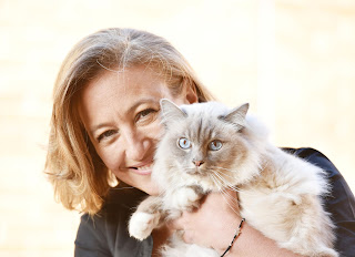
Interview with Pilar Xifra Rubio- Part 1
IODOCAT is the first veterinary practice in Spain that has
ever offered radioiodine treatment for cats with hyperthyroidism. Pilar Xifra
is in charge of this innovative project. In this first part of the interview,
Pilar speaks about how the idea occurred to her and how she put it into
practice. She also talks about her collaboration with Mark Peterson in the book
Feline Endocrinology (2019 Edward C. Feldman, Federico Fracassi, Mark E.
Peterson, Ed Edra S.p.A.) and about different ways to use scintigraphy in veterinary
medicine.
-
Hi Pilar! Thank you for finding time for this
interview. I know you are a busy vet. You have set up the first practice in
Spain that offers radioiodine treatment for hyperthyroid in cats. You also
appear as co-author in Feline Endocrinology. These are really admirable
achievements. Congratulations! Tell us please how did this all happen?!
- Thank
you. It is a pleasure for me to be here. I am now a new fan of
lagateramedicinafelina.blogspot and your podcasts. Regarding my contribution to
the book, I am over the moon. This was
way beyond my expectations. When I received Peterson’s email offering me to
contribute to the chapter about treatment of hyperthyroidism, I just could not
believe it. I had to read the email a few times! I would have never thought I
would be part of such an exciting project.
Regarding IODOCAT it all started at the GEMFE
(Spanish Association of Feline Medicine) Congress in Spain. We were fortunate
to have Peterson here delivering lectures about feline hyperthyroidism. As you
know, I am a member of GEMFE. I am very interested in feline medicine. I could
not understand why we had no radioiodine in Spain. I almost did not know what
they were talking about. It all sounded a bit like Chinese to me. I thought it most likely was very complicated
and that was why we did not have any radioiodine available. Hyperthyroid cats
had to go to Paris if the owners wanted them to be treated. I had a cat with
hyperthyroidism that had adverse effects from methimazole. He developed
pancreatitis amongst other clinical signs and that was not even a curative
treatment. How good is it to be able to cure a disease! And radioiodine allows
you to do that.
A few months after the congress, on a night out, I met a couple of pharmacologist who work at a big hospital here Madrid (Puerta de Hierro). I think that was one of the most productive Gin Tonics I have ever had. One of them is very interested in the veetrinry medicine field. He would have liked to be a vet. We started talking about the lack of radioiodine treatment in Spain and they put me in touch with a nuclear physicist in charge of the nuclear medicine department in Puerta de Hierro.
Compared to what
they do in human medicine he found that helping me would be fairly easy. I
started to regularly go to the hospital to learn about nuclear medicine. I
learnt about scintigraphy and radioiodine. At some point I sent an email to the
doctor or Mark Peterson. I assumed he was not going to reply. At the end of the
day… I am nobody! However he is a very kind man… and he replied! I was so
excited about his email. I also had to read it a couple of times before believing
it. After having a chat with my husband we concluded that I had to go to New
York. I wanted to learn about this somewhere where I could see its application
to veterinary rather than human medicine. Mark Peterson and his team are all
very kind people and I felt really welcome.
It was during
that trip when I made the big decision. I went in March and it was snowing a
lot. Oh my god: for somebody from Spain it is difficult to cope with such a
cold weather when it was meant to be spring! I was not impressed. Anyway, when
I asked Peterson for his opinion about my project he just said: “Done”. So I
came back to Spain and started contacting people I needed help from. That was in
2016. It took me two years to set it all up.
-
Well I think that is not such a long time,
taking into account the magnitude of the project…
-
I guess… But to me it was definitely long! It
was also fun. When I started talking to the nuclear physicist I thought he was
going to think I was crazy: “Really? Treating cats with nuclear medicine?” I
thought that they would laugh at me.
-
How difficult was to process the bureaucratic
side?
-
First I needed a nuclear physicist to help me
getting a license. You also need different devices. We use the gamma camera to
take a visual image of the thyroid tissue. We also have a Geiger counter to
monitor the radiation levels for health and safety purposes. This counter also
helps to work out the dose that we need in every individual case. The person in
charge of the premises needs a special qualification so I had to study quite
hard. I found it interesting but also challenging. There are a lot of technical
terms and physics involved.
-
That sounds challenging indeed. Do you use
scintigraphy for something else besides the treatment of hyperthyroidism in
cats?
-
Initially I only considered its use in feline
hyperthyroidism. However, when I went to the Puerta de Hierro, I started
looking at other possibilities. Actually I have written a paper about the use
of scintigraphy in other pathologies. It will be available next month in AVEPA’s
journal.
-
That is so good! I look forward to reading it!
-
It is a diagnostic method that offers plenty of
advantages compared to other diagnostic tests. It gives you information about the
function of the organ, besides about its anatomical location. An ultrasound or
a CT or an MRI will not offer you that. Another advantage of this diagnostic
test is that the exposure to radiation is much smaller than the exposure
involved in a CT scan or an MRI. CT radiation is 200 times stronger than scintigraphy’s.
Scintigraphy allows you to be with the
patient, we do not need distance in between.
So what is a scintigraphy? This method
involves injecting the patient with a substance containing atoms that emits gamma particles. That allows the gamma camera to catch an image from the
patient's radiation. Besides the radioactive component, this substance that we
inject has also a carrier which “takes” that radioactive component to the
tissues you want to send it to. If you want to see bones you use phosphate as
carrier. That allows you to see where the bone destruction is. We have done
this to diagnose lameness in dogs. Scintigraphy is much more sensitive to detect
this type of lesions than radiography or MRI. It also detects metastasis in
bone tumours at an earlier stage than CT or MRI. We also use it to detect hypothyroidism
in cats and dogs. It is helpful to identify thyroid carcinoma causing hypothyroidism
in dogs with an unidentified mass on the ventral cervical region. It helps to
identify tissue infiltration and metastasis.
Renal scintigraphy is however my favourite
one (after thyroid scintigraphy of course). It is a dynamic scintigraphy, so you
see how the radioactive susbtance is flowing through the kidneys. It allows you
to measure the glomerular filtration rate, what is very helpful before
nephrectomy to assess the function of the remaining kidney. And it detects
kidney injury much earlier than creatinine and SDMA.
-
What's the toxicity associated with this
diagnostic method?
-
There are
no toxic effects.
-
Well… it
is scary to talk about injecting nuclear stuff in cats with renal disease...
-
The radioactive isotope has no harmful effects at
all. It is really useful: it has exclusively glomerular filtration and no
tubular filtration. In human medicine there are no boundaries diagnostic tests.
They frequently use scintigraphy and also PET- CT, which is also nuclear
medicine. For instance, by marking neutrophils and injecting them back into the
patient we can find the specific location of a hiding infections, sites of
inflammation or gastrointestinal ulceration and haemorrhage.
-
Wow it sounds like science fiction!
-
We also use it to detect portosystemic shunt
(PSS). It is a very simple test where the pertechnetate is administered as a
colonic enema. In a normal cat it would go first to the liver and then to the
heart. If it he goes straight to the heart without going to the liver first, there
is a PSS. It allows you to distinguish an extra hepatic from an intra-hepatic shunt.
However it is not as sensitive as a CT scan at locating the intra hepatic
shunt. I still find it very useful in situations where the practitioner is not
sure about the presence of PSS or in
cases with new colateral PSS. It is less invasive than other advanced imaging
tests, it does not require general anaesthesia and the amount of radiation exposure
is also reduced. If the cats is cooperative and stays still for a few minutes whilst
you inject the drug, it does not need sedation.
Bone scintigraphies are the most time-consuming
once because the patients are usually big dogs. It may take a few hours to get it
done.
-
And what type of substance do you use in these
cases?
-
The main substance used in scintigraphy is technetium,
usually combined with something else. In bone scintigraphy we use Technetium
HDP, whereas in renal scintigraphy we use Technetium DTPA. They need acronyms
because they all have different and very complicated and long names! In liver
scintigraphy we use pertechnetate, the same as in thyroid scintigraphy. The
half-life of a radioactive isotope is the time it takes for its effect to drop
to 50% of its initial value. Technetium has a half-life of only 6 hours and generates
a much smaller radiation than I131, which has a half-life of 8 days. That is
why the patient remains radioactive for a longer time. In human medicine the
most used iodine is I123, which has an even shorter half-life and does not
destroy thyroid tissue. The aim is to minimise the side effects. I131 would
allow us to locate the abnormal thyroid tissue, but we would be treating it at
the same time and that would generate a greater amount of radiation that we can
avoid by using pertechnetate instead…
The second part of this interview will be available in a few
days….

